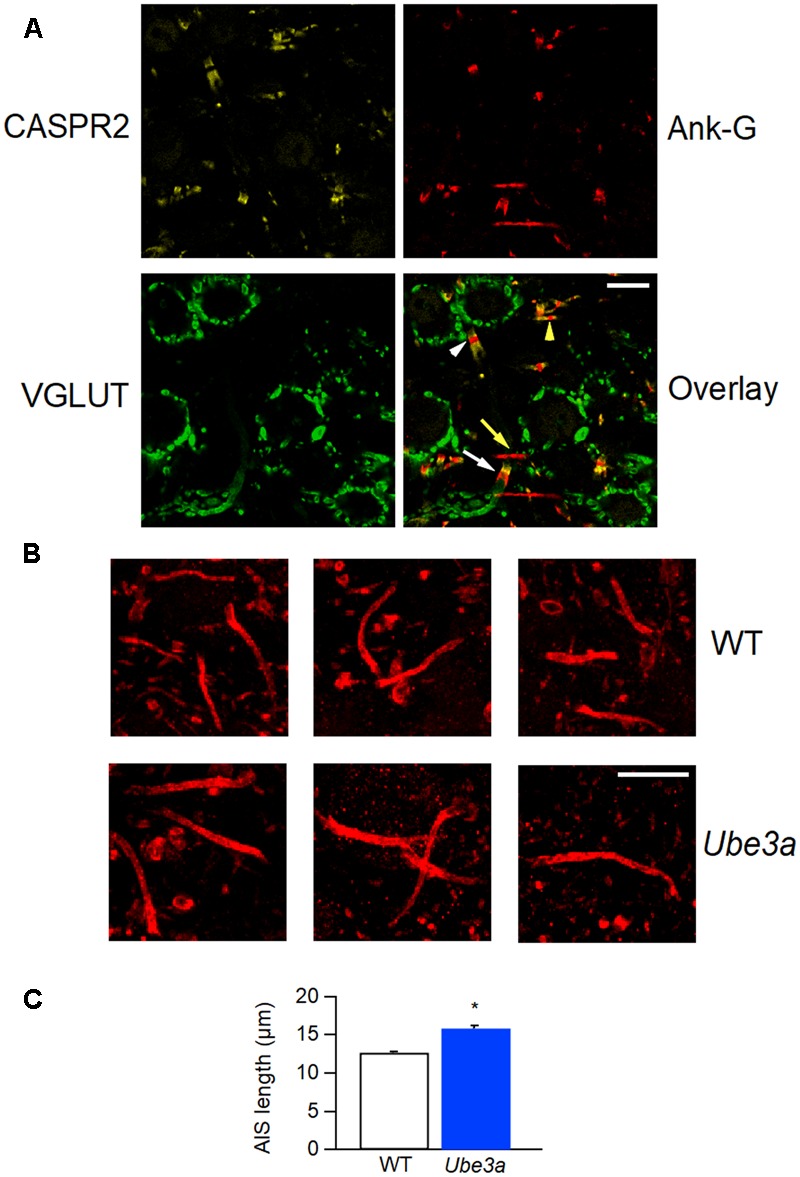
FIGURE 11.

Longer AIS in Ube3a mice. (A) Confocal section of MNTB slice, labeled with anti-CASPR2 (yellow) and anti-ankyrin-G (red) and anti-Vglut1 & 2 (green). The composite image shows a node of Ranvier from a calyceal axon (white arrow head) and a principal cell axon (yellow arrow head), and a calyceal heminode (white arrow), as well as a postsynaptic AIS (yellow arrow). (B) AISs of MNTB cells labeled by anti-ankyrin-G, from both WT and Ube3a animals. (C) Comparison of AIS length in C57BL/6J WT and Ube3aE113X mice. ∗Indicates p < 0.001. Calibration bars 10 μm.
