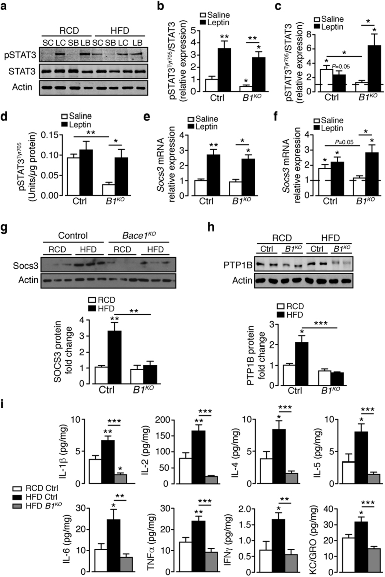
Figure 3.
Absence of Bace1 enhances leptin signalling in DIO mice and prevents HFD-induced hypothalamic inflammation. (a) Immunoblots of total STAT3, actin and pSTAT3Tyr705 levels in saline- and leptin-stimulated control (SC, LC) and Bace1 KO (SB, LB) mice on RCD or HFD. Ratio of signal intensities for pSTAT3Tyr705 to STAT3 in RCD (b) and HFD (c) mice (n = 6–9/group). (d) pSTAT3 by ELISA for RCD and HFD control (n = 7) and Bace1 KO (n = 8) mice. (e,f) RT-qPCR for Socs3 from saline and leptin-treated control and Bace1 KO mice (n = 6–10/group) on RCD (e) and HFD (f). (g,h) Immunoblots of hypothalamic SOCS3 (g) and PTP1B (h) and actin of control and Bace1 KO mice on RCD and HFD. Quantified signal intensity of SOCS3 (n = 3–8/group) and PTP1B (n = 6–13/group) to actin. (i) Hypothalamic cytokine levels for RCD and HFD control and HFD Bace1 KO mice (n = 11–14/group). Data are means ± SEM. *P < 0.05, **P < 0.01, P < 0.001 by Kruskal-Wallis test (b,c,e,f,h) or one-way ANOVA with Bonferroni’s multiple comparisons test (d,i). Uncropped images of immunoblots can be found in Supplementary Fig. 10.

