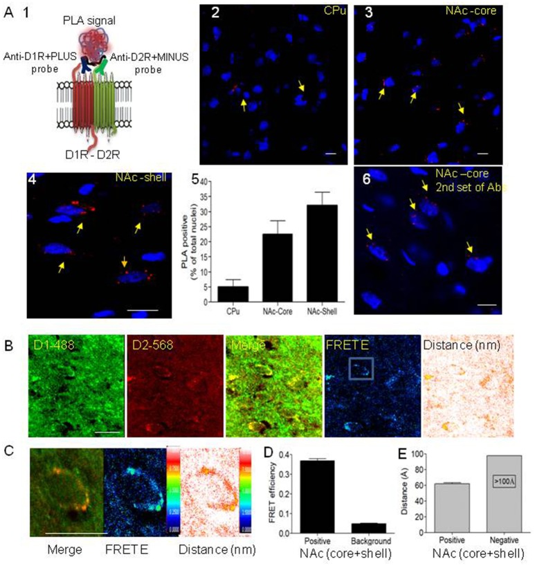
Figure 1.
Evidence for the existence of dopamine D1-D2 receptor heteromer in rat NAc. (A) Proximity ligation assay (PLA) was used to visualize and detect D1R and D2R close proximity. (A1) A scheme depicts the PLA probes used in the present study. (A2–A4) Representative images of PLA signals (red dots) in neurons (nuclei stained by DAPI) in rat caudate putamen (CPu), nucleus accumbens core (NAc-core) and shell (NAc-shell) subregions. (A5) Graph representing the percent of neurons with a positive PLA signal. (A6) Representative image of PLA signals in neurons (nuclei stained by DAPI) in rat NAc-core using the second set of antibodies. (B) Representative images of immunohistochemistry using D1R antibody (D1R-Ab) or D2R antibody (D2R-Ab) directly conjugated to Alexa-488 or Alexa-568, respectively, in the NAc-shell. Direct confocal FRET analysis was performed, reflected by FRET efficiency (FRET E) and the distance between the dipoles, less than 10 nm (100 Å). (C) A representative close-up of a single MSN cell body from NAc showing D1R-D2R colocalization (left), D1-D2 heteromer FRET efficiency (center) and relative distance between receptors (right). (D,E) Histograms showing FRET E ratios (D) and distance (E) obtained from MSN cell bodies from NAc (n = 24). Bars are 10 μm.

