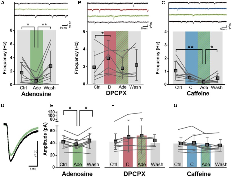
FIGURE 3.

A1R-mediated effects of adenosine on synaptic transmission are post-synaptically blocked by caffeine. (A) Adenosine (20 μM, green) significantly decreased the frequency of excitatory events onto pyramidal neurons in a reversible manner. Paired t-test comparing adenosine (20 μM) to ACSF condition: ∗p < 0.05; ∗∗p < 0.01. Boxes depict average ± SD. (B) Direct application of DPCPX (100 nM, red) significantly increased the frequency of excitatory events and blocked adenosine-induced inhibition (green). Paired t-test comparing DPCPX (100 nM) to ACSF condition: ∗p < 0.05. Boxes depict average ± SD. (C) Caffeine pre-incubation (50 μM, blue) did not affect the potency of adenosine (20 μM, green) to decrease the frequency of excitatory events onto pyramidal neurons. Wash-out of adenosine and caffeine restored the frequency of events. Paired t-test comparing adenosine (20 μM) to ACSF condition: ∗p < 0.05; ∗∗p < 0.01. Boxes depict average ± SD. (D) Example trace of the average effect of adenosine application onto the amplitude of excitatory events onto a pyramidal neuron. (E–G) Adenosine (20 μM, green) significantly decreased the amplitude of excitatory events onto pyramidal neurons in a reversible manner (E). Pre-incubation with either DPCPX (100 nM, red, F) or caffeine (50 μM, blue, G) completely abolished this effect. Paired t-test comparing adenosine (20 μM) to ACSF control/wash-out condition: ∗p < 0.05.
