Figure 7.
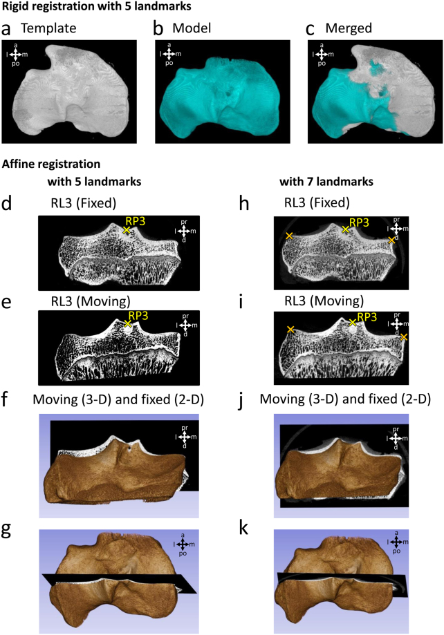
Assessment of the usability of the reference points for landmark-based 3-D registration. (a–c) Landmark-based rigid registration of two tibial plateau samples in the Fiji software, using RP1-5 as landmarks. 3-D view of the (a) template and (b) model image after registration. (c) Merged view of the template and model images. Landmark-based affine registration of the same two samples in the 3D Slicer software, (d–g) using only RP1-5 as landmarks, and (h–k) using additional 2 landmarks (labelled with orange crosses) to RP1-5, placed on the medial end of the medial tibial plateau and the lateral end of the lateral tibial plateau on the 2-D section corresponding to RL3. Images show the transformed samples after registration. Example micro-CT images corresponding to RL3 of the samples set as the (d,h) fixed and (e,i) the moving volume. (f,j) Posterior and (g,k) top view of the two samples merged after registration. The 3-D volume shows the sample set as the moving volume, and the 2-D micro-CT section shows RL3 of the sample set as the fixed volume. Abbreviations: a: anterior, d: distal, l: lateral, m: medial, pr: proximal, po: posterior.

