Figure 5.

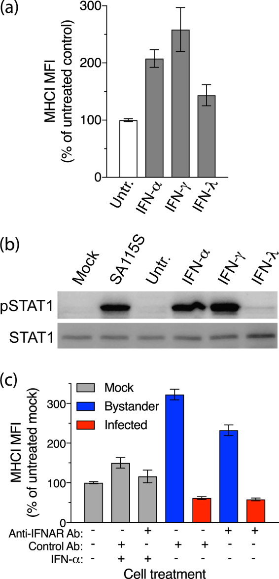
Involvement of IFN receptor signalling in modulation of MHCI expression by rotavirus. (a) Cells left untreated (Untr.) or treated with IFN-α, IFN-γ or IFN-λ for 16 h were fixed and stained for total MHCI and analysed by flow cytometry. (b) Cells were mock-infected (Mock) or infected with SA11-5S at a m.o.i of 1 for 16 h, or left untreated or treated with IFNs as for (a), and analysed by Western blot for levels of STAT1 or phosphorylated STAT1 (pSTAT1). Full-length blots are presented in Supplementary Figure 3. (c) Cells were mock-infected or infected with SA11-5S at a m.o.i. of 1 in the presence of IFN-α and/or blocking antibodies to IFNAR1, or control antibodies. After 16 h cells were fixed and permeabilized, stained for MHCI and rotavirus, and analysed by flow cytometry. For (a,c), the mean ± S.D. of the MHCI MFI from three biological replicates is shown.
