
Figure 5.
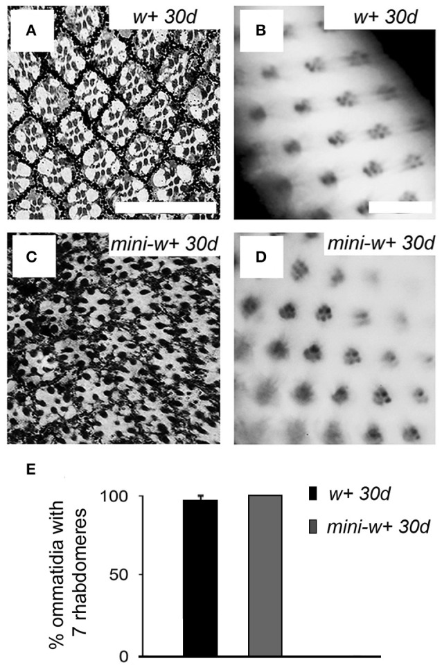
Expression of the mini-white+ transgene in w1118 mutants rescues the retinal degeneration phenotype. Histological microscopy sections of the retina from 30 days-old w+ flies from the Vallecas strain (A) and transgenic flies expressing mini-w+ in a w1118 null genetic background (C) showed a w+-like organization as neither missing ommatidia nor rhabdomeres was observed (compare with w− mutants, Figures 1F,G). (B,D) Images of the retina obtained by the method of optical neutralization of the cornea in w+ (B) and transgenic flies expressing mini-w+ (D), showing that the expression of mini-w+ in a w− background rescues the phenotype. Scale bars represent 20 μm. (E) Graphical representation of the percentage of ommatidia with seven rhabdomeres in 30 days-old flies from w+ and mini-w+ strains showing that there are no significant differences in this parameter between both genotypes (w+ 30 d vs. mini-w+ 30 d, Mann–Whitney U-test, p >0.05).