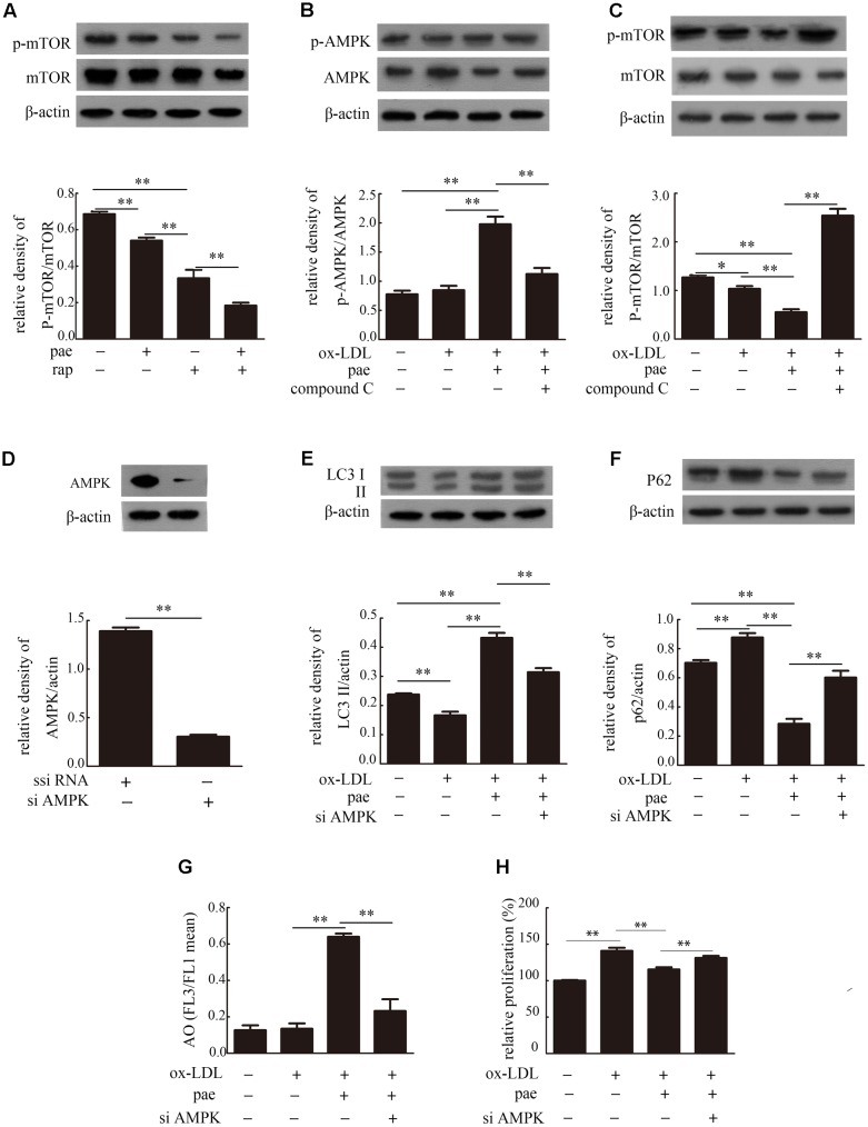
FIGURE 7.
Paeonol induced autophagy in VSMCs was via the AMPK/mTOR signaling pathway. (A) Representative blots of p-mTOR/mTOR in VSMCs treated with Paeonol in the absence or presence of rap. Representative blots of p-AMPK/AMPK (B) and p-mTOR/mTOR (C) in ox-LDL-induced VSMCs treated with Paeonol in the absence or presence of compound C. (D) Immunoblot verification of AMPK knockdown. Representative blots of LC3II (E) and p62 (F) in VSMCs or si-AMPK transfected cells treated with ox-LDL and Paeonol. (G) Intracellular acidification was calculated by red-to-green (FL3/FL1) mean fluorescence intensity. (H) VSMC proliferation was analyzed by a CCK8 assay. Data are the mean ± SD, n = 3. ∗p < 0.05, ∗∗p < 0.01.

