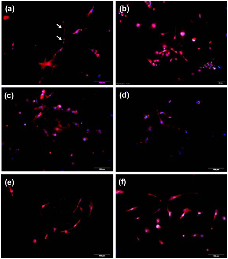
Figure 6.
Immunofluorescence staining for the expression of the (a) synapsin I (red), (b) vesicular glutamate transporter 2 (VGlut2, red), (c) γ-Aminobutyric acid (GABA, red, (d) Tau protein (red), (e) tyrosine hydroxylase (Th, red), and (f) neuronal nuclei (NeuN, red) under the treatment of 256 µg/mL pABM-I@M for 19 days. The MFs were transfected for 1 hr) and the nuclei were counterstained with 4′,6-diamidino-2-phenylindole (DAPI, blue). Scale bars are 100 μm.

