Figure 1.
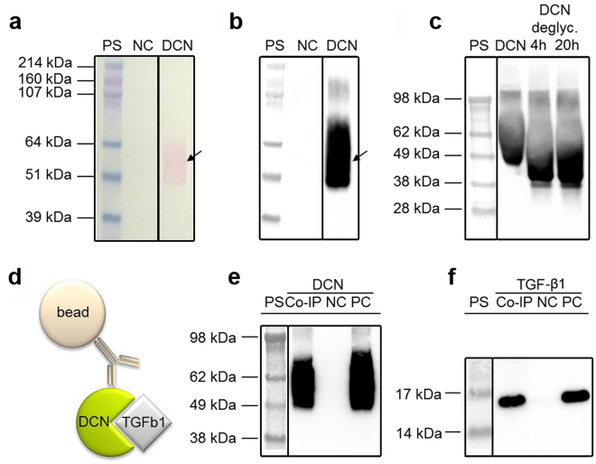
Characterization of recombinant human DCN. (a) Ponceau-Red staining of a nitrocellulose membrane containing an IMAC purification of suspension media with 1% dialyzed FBS (negative control, NC), an IMAC purification of DCN-containing cell culture media with originally 1% dialyzed FBS (DCN), and the HiMark Pre-stained protein standard (PS). (b) The same membrane after discoloring and specific immunodetection of DCN. (c) Specific immunodetection of untreated DCN, deglycosylated DCN (DCN deglyc., 4 h and 20 h) and the SeeBlue® Plus2 Pre-stained protein standard (PS). (d) Schematic of a Co-IP for the protein interaction between DCN and TGF-β1. (e,f) Specific immunodetection of human DCN (e) or TGF-β1 from human platelets (f) show the Co-IP eluate (Co-IP), an unspecific background control without the DCN antibody (NC), the positive control (PC; 250 ng DCN or 100 ng TGF-β1) and the SeeBlue® Plus2 Pre-stained protein standard (PS). All blots are cropped, indicated by the black boxes. Full-length blots are presented in Supplementary Figure 7.

