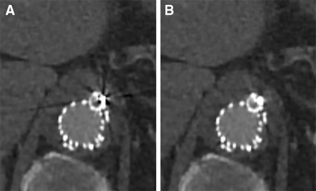
Fig. 1.

CT images (A standard (AIDR 3DE), B SEMAR) of a branch connected to the celiac artery in a 65-year-old male. Artifacts (black streaks) are reduced on the CT image with the SEMAR reconstruction compared to the standard reconstruction. All CT images are displayed with a window level of 300 HU and a window width of 1000 HU
