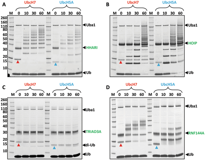
Figure 3.
Auto-ubiquitination assays of RBRs with UbcH7 and UbcH5A. Coomassie-stained reducing SDS-gels of auto-ubiquitination assays for HHARI-RBR (A), HOIP-RBR (B), TRIAD3A-RBR (C) and RNF144A-RBR (D). The concentration of RBR used was 5 μM, whilst the concentrations of E1, E2 and Ub were 0.1, 2 and 50 μM, respectively. 10 mM ATP was added to start the reaction and samples were taken after 10, 30 and 60 minutes incubation at room temperature. Red and blue arrowheads indicate UbcH7 and UbcH5A, respectively; black arrowheads indicate E1 (Uba1), E3 and ubiquitin (Ub). In the case of TRIAD3A accumulation of di-ubiquitin (di-Ub) is observed, indicated by a black arrowhead.

