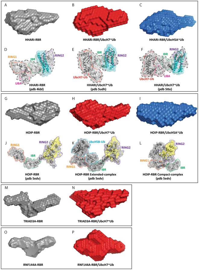
Figure 5.
SAXS-derived envelopes and comparison with available crystal structures of HOIP/UbcH5B-Ub and HHARI/UbcH7-Ub. Low resolution SAXS-derived molecular envelopes for isolated RBRs domains (A,G,M and O) and their complexes with UbcH7-Ub and UbcH5A-Ub (B,C,H,I,N and P). Crystal structures of HHARI (PDB 4KBL) and HHARI/UbcH7-Ub (PDB 5UDH and 5TTE) are reported in panel D, E and F, respectively. The domains that constitute the RBR and E2-Ub are circled by dashed coloured lines, with UBA in pink, RING1 in orange, IBR in green, RING2 in purple and UbcH7-Ub in red. The Ariadne domain, missing in the construct used for SAXS analysis, is shown in cyan. A black dashed line contours the portions of the structures that span the same number of domains as in the construct used in our SAXS analysis. The structures of the RBR domain of HOIP, and the extended- and compact- conformations in presence of UbcH5B-Ub, derived from PDB 5EDV, are reported in (J–L). The RBR domains and the E2-Ub are circled by dashed coloured lines, with RING1 in orange, IBR in green, RING2 in purple and UbcH5B-Ub in blue. In the 5EDV structure, the RING2 of HOIP is poorly defined; therefore we have reconstructed the missing regions in yellow using the higher resolution crystal structure of RING2 reported in PDB 4LJQ.

