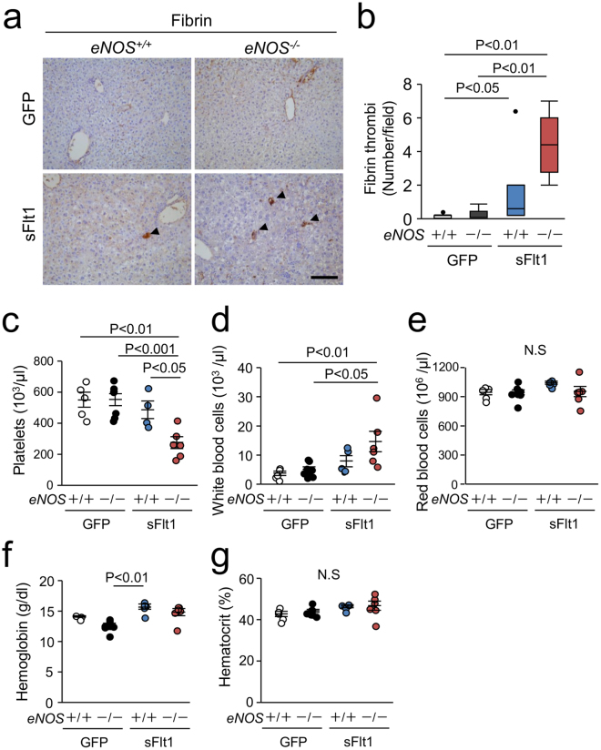
Figure 6.
Fibrin deposition in the liver and thrombocytopenia. (a) Representative photomicrographs of immunohistochemistry against Fibrin. Scale bar indicates 100 µm. (b) Number of Fibrin thrombi is significantly increased in the liver from eNOS −/−; sFlt1 mice. (c–g) Data of blood count; platelets (c), white blood cells (d), red blood cells (e), hemoglobin (f), and hematocrit (g). Excessive sFlt1 combined eNOS deletion causes thrombocytopenia. N.S., not significant. n = 4–8. Data are shown as mean ± s.e.m or box plot. ANOVA or Kruskal-Wallis test.

