Figure 5.
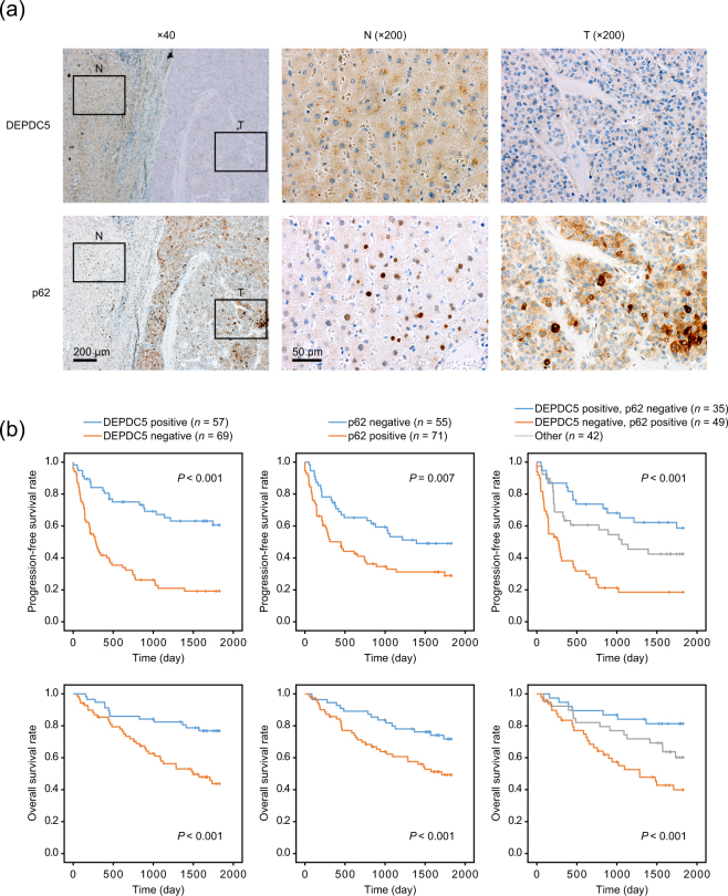
Relationship among DEPDC5 and p62 expression in HCC samples and patient prognosis. (a) Immunohistochemical analysis of DEPDC5 and p62 in a representative tissue sample including adjacent liver tissue (N) and cancer (T). Nuclei were counterstained with hematoxylin. In adjacent liver tissues of almost all cases, DEPDC5 was positive while p62 was negative. (b) Kaplan-Meier curves of the progression-free and overall survival in groups of HCC patients classified according to DEPDC5 and p62 expression patterns. P values were calculated by the log-rank test.

