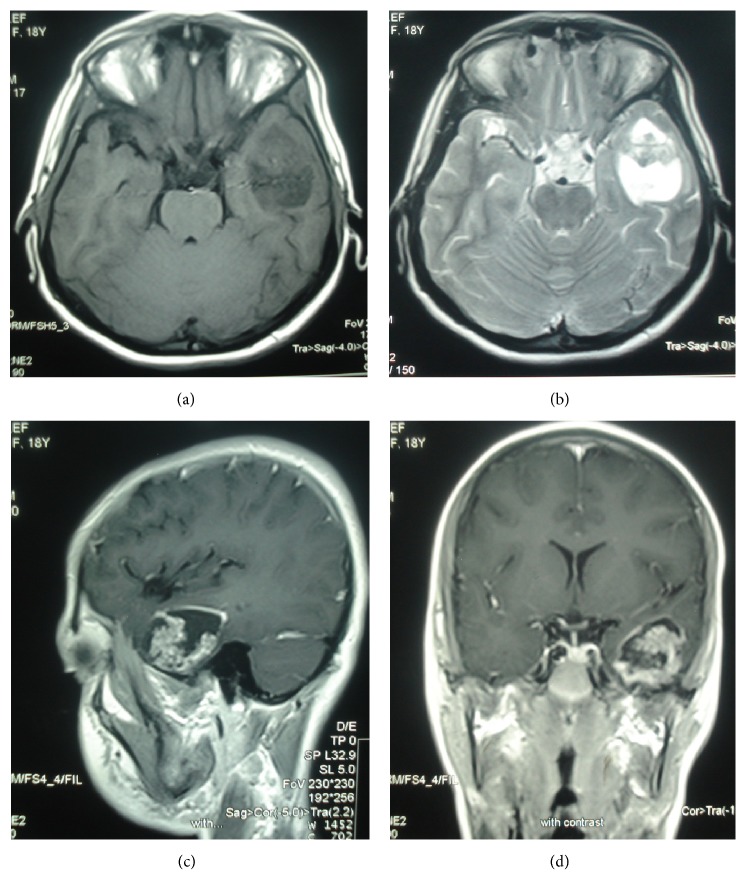
Figure 1.
Craniopharyngioma of the left temporal lobe. Axial T1-weighted (a) and T2-weighted (b) MR images show a solid-cystic mass with no significant edema at left temporal lobe. Sagittal (c) and coronal contrast enhanced T1-weighted (d) MR images show peripheral wall enhancement and enhancing solid component.

