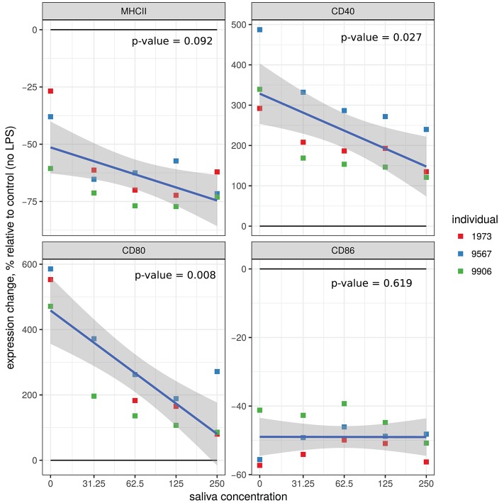
Figure 2.
Effect of A. variegatum saliva on the expression of surface markers on LPS-stimulated bovine macrophages. Bovine blood monocyte-derived macrophages were pre-stimulated for 1 h with LPS, and then for an additional 24 h with different concentrations of A. variegatum saliva. Cells were collected and stained for surface markers. Expression levels (mean of fluorescence intensity, MFI) of MHC II, CD40, CD80, and CD86 markers on macrophages compared with unstimulated control cells were measured by flow cytometry. Data are means of triplicate experiments, for a pool of saliva batches for the three animals tested. Blue lines and shading show the linear regression fit and the 0.95 confidence interval, respectively. The p-value of the model is indicated on the panel.

