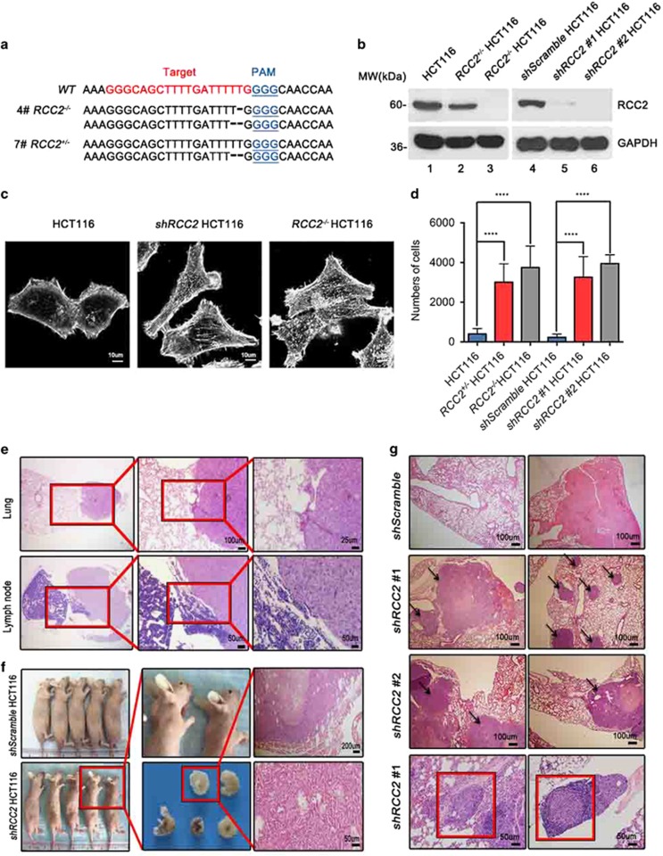
Figure 2.
Reduction of RCC2 facilitates cell migration and tumor metastasis. (a) Generation of CRISPR-mediated RCC2 knockout cell lines. Target site and PAM motif are illustrated with red and blue. Mutations of number 4 and 7 clones are shown with specific nucleotide deletions. (b) Elimination of RCC2 protein expression in shRCC2 #1, shRCC2 #2, RCC2+/− and RCC2−/−cells. (c) Morphology of HCT116, shRCC2 and RCC2−/− HCT116 cells shown with phalloidin staining. Cells were scanned with confocal microscopy. (d) Average numbers of migrated HCT116, RCC2+/−, RCC2−/−, shScramble, shRCC2 #1 and shRCC2 #2 HCT116 cells were calculated using the transwell assay. All data are presented as mean±s.e.m. and were analyzed by the unpaired t-test (n=4). ****P<0.0001. (e) Hematoxylin and eosin (H&E)-stained tissues from nude mice inoculated with shScramble and shRCC2 cells. Metastases found in lung tissues and lymph nodes are shown at various magnifications. Red boxes indicate the margins of normal tissue (left) and tumors (right). (f) Of the nude mice intravenously injected with shRCC2 cells, 40% produced tumors of the back or neck compared with the control group injected with shScramble cells. Each group contained five individual nude mice. (g) H&E staining shows shRCC2 cell metastases to lung tissue with formation of multiple tumor colonies in nude mice, but shScramble cells caused only necrosis. Red boxes show cells invading a blood vessel (left) and a lymph node (right), and arrows indicate metastatic tumor. See also Supplementary Figure S2.

