Figure 4.
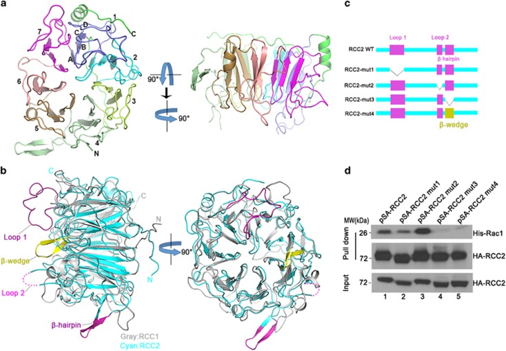
Overall structure of RCC2 and the structural elements accounting for interaction with Rac1. (a) Ribbon diagram of the RCC2 propeller structure viewed along or perpendicular to the central shaft. The blades are numbered (1–7) along the sequence. (b) Superimposition of human RCC2 (cyan) and human RCC1 (gray) structures viewed perpendicular to or along the central shaft. The extra structural elements in RCC2 (loop 1, loop 2 and β-hairpin) relative to RCC1 are shown in magenta. The extra β-wedge required for RCC1 guanine exchange factor activity is shown in yellow. (c) RCC2 deletion mutants used in this study. (d) S-tag-HA-tag RCC2 mutants were expressed in vivo and lysates were incubated with purified His-Rac1 protein. Rac1 or RCC2 was then evaluated with anti-Rac1 or anti-HA antibody respectively. See also Supplementary Figure S4.

