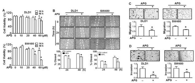
Fig. 2.
Apigenin inhibits cell migration and invasion of DLD1 and SW480 cells. (A) DLD1 and SW480 cells were treated with various doses of apigenin (APG) or vehicle (DMSO) for 24 or 48 h. Cell viability was determined by CellTiter 96® cell proliferation assay. * p < 0.05 compared to control without treatment. (B) Representative images of wounded DLD1 cells and SW480 cells treated with or without 20 μM APG for 24 and 48 h. Cell migration was determined by percentage of closure of wound gap at time 0. * p < 0.05 compared to control without treatment at corresponding treatment time. (C) and (D) Cell migration and invasion determined by Boyden chamber assay. Representative images of cell migration (C) and invasion (D) in DLD1 cells and SW480 cells with or without 20 μM of APG treatment. All values were expressed as fold changes relative to none treatment control. * p < 0.05 compared to none treatment control.

