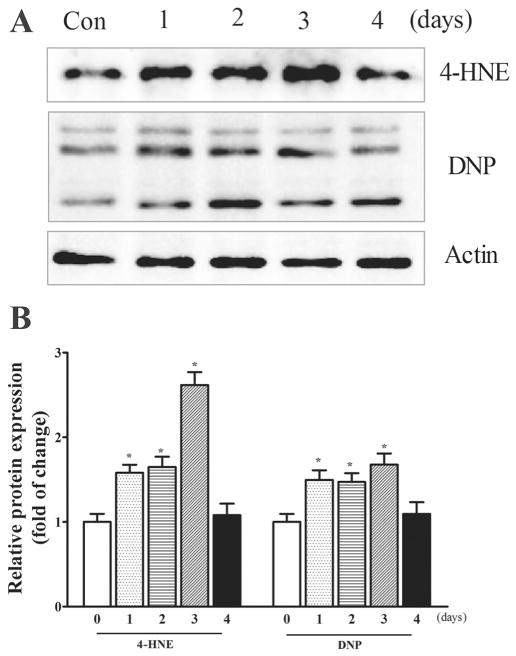
Fig. 3.
TD-induced oxidative stress in iCell Neurons. A. iCell neurons were treated with amprolium (0 or 1 mM) At specified times after the treatment, the lysates of iCell neurons samples were collected. The expressions of 4-HNE and DNP were determined by immunoblotting. B. The relative amounts of 4-HNE and DNP were measured microdensitometrically and normalized to the expression of actin. The experiment was replicated three times, and the results were expressed as the mean ± SEM. *p < 0.05, statistically significant difference from untreated control groups.

