Fig. 2.

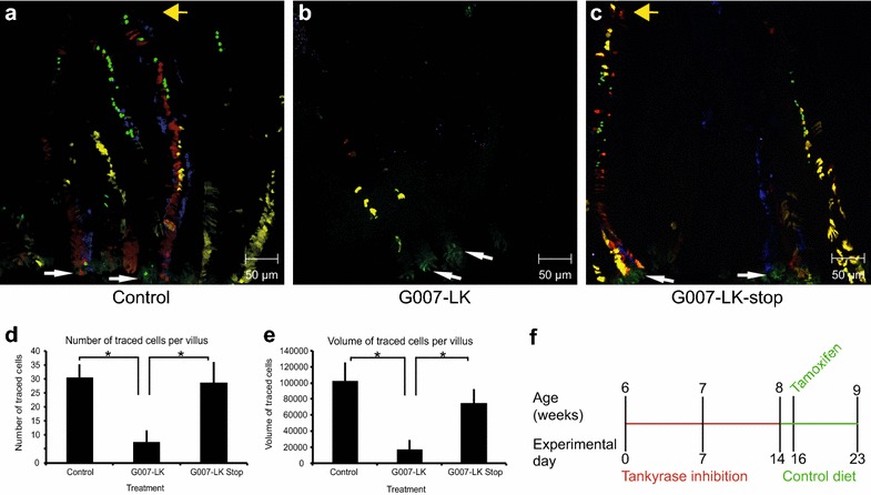
Lineage tracing from LGR5+ cells in the duodenum. a, b Confocal imaging of duodenal tissue in a Lgr5-EGFP-Ires-CreERT2;R26R-Confetti mouse after 7 days of lineage tracing in control (a) and G007-LK-treated, enriched chow 100 mg G007-LK/kg (b) mice according to the G007-LK administration schema outlined in Fig. 1f. LGR5+ stem cells express EGFP in the cytoplasm and are identifiable at the crypt base (white arrows indicate LGR5+ cells at the crypt base, yellow arrows indicate the tip of the villi). Traced LGR5+ cell progeny express RFP (red fluorescent protein), YFP (yellow fluorescent protein), EGFP (in the nucleus) or CFP (cyan fluorescent protein). c Confocal imaging of duodenal tissue in a Lgr5-EGFP-Ires-CreERT2;R26R-Confetti mouse after termination of G007-LK treatment (enriched chow, 100 mg G007-LK/kg chow, G007-LK-stop) prior to initiation of LGR5+ cell progeny tracing (administration schema outlined in f). d Quantification of the number of traced cells in Control, G007-LK and G007-LK-stop treated mice (mean ± SEM, *p < 0.05, n ≥ 3). e Quantification of the volume of traced cells in Control, G007-LK and G007-LK-stop treated mice (mean ± SEM, *p < 0.05, n ≥ 3). f Administration schema for G007-LK enriched chow in the G007-LK-stop group, G007-LK administration was stopped 2 days prior to initiation of lineage tracing by IP injection of tamoxifen
