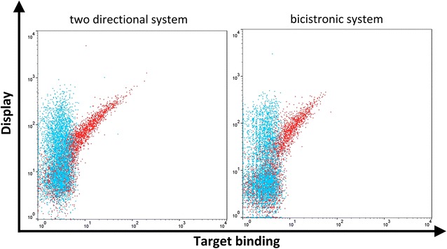
Fig. 2.

Overlay of trastuzumab displaying yeast cells either stained with detection antibodies only (blue) or with detection antibodies and HER2 as monitored by flow cytometry. Yeast cells were consecutively incubated with 1 µM of His tagged HER2 followed by secondary labeling with Alexa Fluor 647 conjugated anti-Penta-His antibody (target binding) and PE conjugated anti-kappa-antibody (display)
