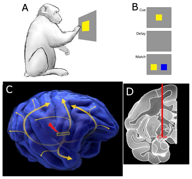
Figure 1. Task paradigm and electrical stimulation position in the brain.
A. Macaque monkeys were trained to interact with a touchscreen. The first step in the task is touching the cue. B. After the cue is touched, the screen blanks during the delay period, followed by presentation of two potential matches. Three colors were used in training, and two were randomly selected as cue/match and distractor on each trial. C. Stimulation of the Nucleus Basalis of Meynert was used in experimental sessions. The position of the nucleus is shown by the dotted yellow rectangle, with the curved arrows approximating the pathways from the nucleus to neocortices[52]. D. An MRI coronal section of a Rhesus monkey brain with the implantation target outlined in red. The MRI was taken from the Macaque Scalable Brain Atlas[53,54]. See also Figure S1–S2.

