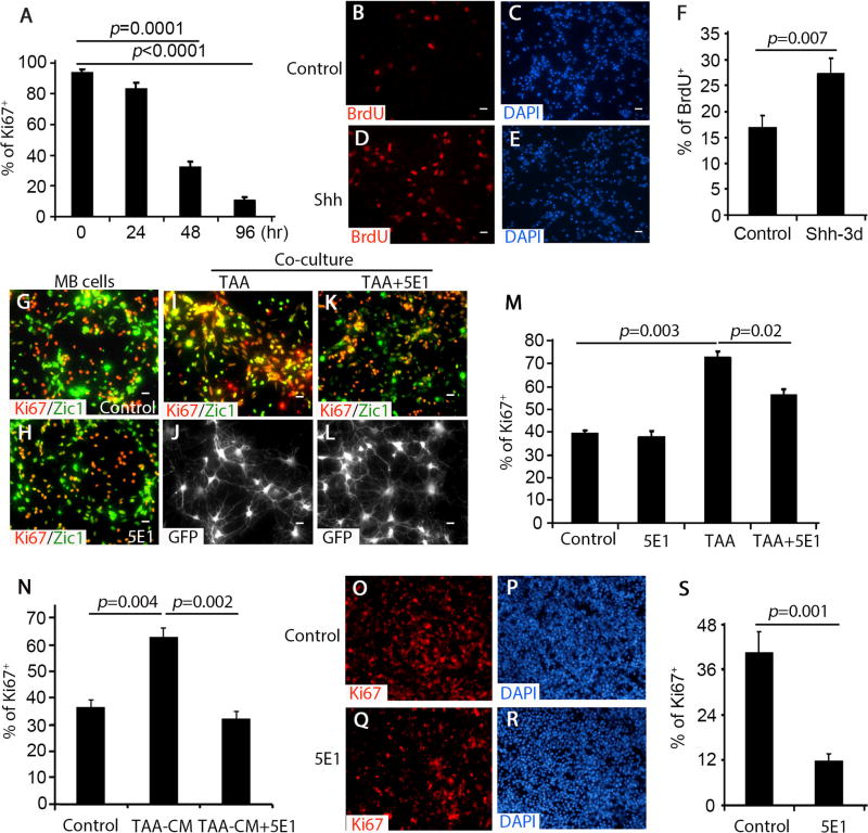
Figure 3. TAA-derived Shh supports MB cell proliferation.
A, Percentage of Ki67+ cells among cultured MB cells harvested at designated time points (0–96 hrs) was quantified. B–E, MB cells were treated with PBS control (B and C) or Shh (D and E) for 3 days, and immunostained for BrdU after a 2 hour pulse with 100µM BrdU. F, The percentage of BrdU+ cells among control (DMSO) or Shh-treated MB cells was quantified. G–N, MB cells cultured alone (G and H) or co-cultured with TAA (I and J) together with 1% 5E1 (K and L) were immunostained for Ki67, Zic1 or GFP. MB cells were treated with NB-B27 (control), TAA-CM, or together with 5E1 for 48hrs and immunostained for Ki67 (N). The percentage of Ki67+ cells among cultured MB cells was quantified in M and N. O–S, MB slices were immunostained for Ki67 after being culture without treatment (O and P) or 5E1 (Q and R) for 4 days. The percentage of Ki67+ cells among MB cells in MB slices was quantified (S). DAPI was used to counterstain cell nuclei in C, E, O and Q. Data in figure A, F, M, N and S represent means ± SEM from three independent experiments. Scale bar, 10 µm.

