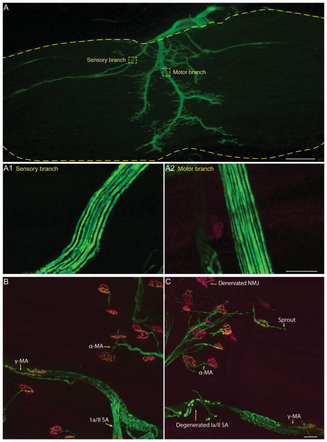
Figure 3.
Visualizing sensory and motor axons in the EDL muscle. Most nerve endings can be fully visualized in whole-mounted EDL muscles from mice expressing YFP (A). Sensory (A1) and motor (A2) branches can be traced from their nerve endings and counted. The nerve endings are shown in control (B) and SOD1G93A (C) animals. α-MA = α-motor neuron axon; γ-MA = γ-motor neuron axon; Ia/II SA = Ia/II sensory afferent; NMJ, neuromuscular junction.

