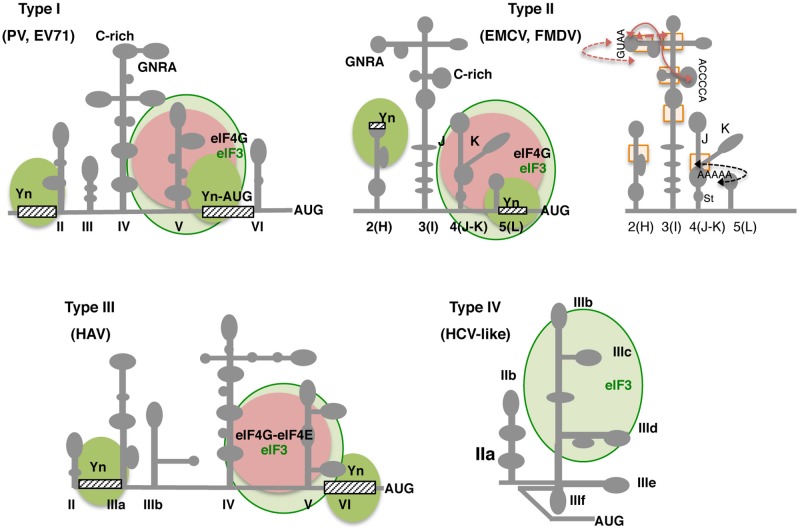
FIGURE 4.
Secondary structure and conserved RNA motifs of picornavirus IRES elements classified as types I, II, III, and IV. Representative members of type I are PV and EV71, members of type II are EMCV and FMDV. Type III is present in HAV, and type IV, also termed HCV-like, is represented by teschovirus IRES. Gray lines depict IRES domains: II–VI in type I; 2 to 5 (or H to L) in type II; II–VI in type III, and IIa to IIIf including a pseudoknot (PK) in type IV. Conserved motifs (GNRA, C-rich, Yn, A-rich) are indicated. Approximate binding sites of eIF3 in all types (light green circle), eIF4G in types I and II, or eIF4G-eIF4E complex in type III (pink circle) are indicated; the recognition of the pyrimidine-rich Yn motif by the polypyrimidine binding protein (PTB) is depicted by solid green circles. Other factors interacting with the IRES are mentioned in the text. For type II IRES elements, brown arrows denote tertiary interactions within the apical region of domain 3; a black arrow depicts the rearrangement of subdomains J-K and stem (St) of domain 4 as a consequence of eIF4G binding (right panel). Rectangles denote regions of local flexibility identified by RNA footprint using dimetallic compounds.

