
FIGURE 5.
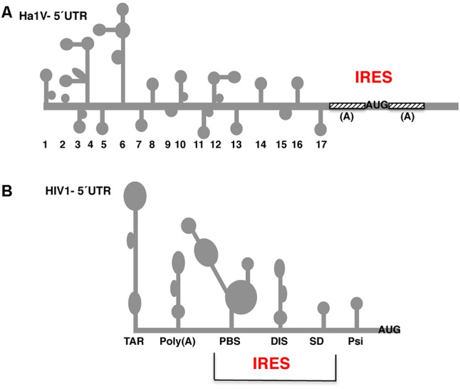
Secondary structure of the 5′ UTR of Halastavi arva virus (HaIV) and HIV-1 RNAs. (A) Stem-loops (1–17) of HaIV 5′ UTR are indicated; A-rich unstructured regions flank the functional AUG codon. (B) Structural motifs of the HIV-1 5′ UTR are schematically represented. The minimal IRES element overlaps the primer binding site (PBS), dimerization initiation site (DIS), and splice donor (SD) stem-loops. RNA structural motifs within the 5′ UTR flanking the minimal IRES are the trans-activating region (TAR) and the polyadenylation signal (PBS) at the 5′ end, and the packaging signal (Psi) downstream of the IRES element.