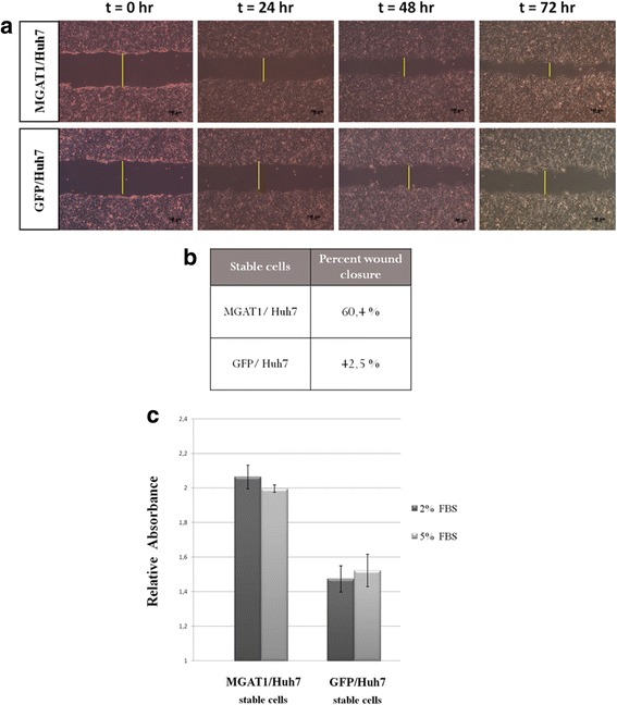
Fig. 4.

Wound healing assay for the stable cells overexpressing the MGAT1 and GFP (control) genes. The bright field images are representative of two independent experiments (a). The distances across the wound are measured by Image J and the differences are presented as percent wound closure (b). MGAT1 stable expression in Huh7 cells results in higher proliferation rate compared to GFP-expressing control stable cells, as determined by XTT assay. The graph indicates results from two independent experiments (c)
