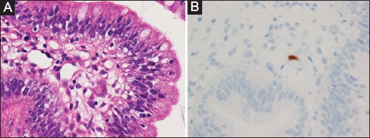
Figure 2.

Photomicrographs of the biopsy specimen taken from the major papilla. (A) A very small number of cytomegalovirus inclusions were seen (hematoxylin and eosin stain; original magnification, 400×). (B) The cells with cytomegalovirus inclusions showing positive immunoreactions to the presence of cytomegalovirus (original magnification, 400×)
