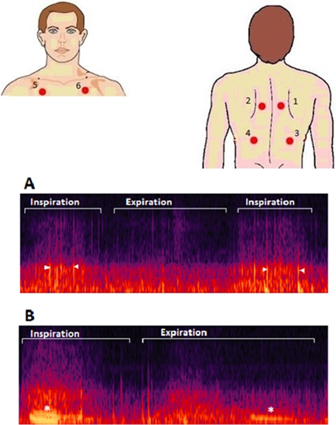
Figure 1.

Upper: illustration showing the different places where lung sounds were recorded. (1_2) Between the spine and the medial border of the scapula at the level of T4–T5; (3_4) at the middle point between the spine and the mid-axillary line at the level of T9–T10; (5_6) at the intersection of the mid-clavicular line and second intercostal space. Lower: image showing two different spectrograms containing crackles (A) and wheezes (B). Crackles appear as vertical lines (arrowheads) and wheezes as horizontal lines (*).
