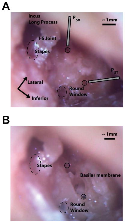
Fig. 1.
Photomicrographs showing the placement of the intracochlear pressure probes in the scala vestibuli and scala tympani before (A), and after (B) removing the bone between the two cochleostomies in specimen 55R (right ear). Dashed circles indicate the approximate locations of the oval and round windows (not to scale); solid circles indicate the entry points of the pressure probes into the cochlea. Note the glass beads located on the stapes capitulum and stapedius muscle/tendon in A, and the location and orientation of the basilar membrane in B. Additional labels are located on the incus long process, incudostapedial (I-S) joint, stapes, round window, and basilar membrane, in addition to the approximate orientation of the pressure probes entering scala vestibuli (PSV) and scala tympani (PST).

