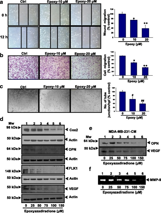
Fig. 5.

Epoxyazadiradione inhibits cell migration, HUVECs tube formation and pro-angiogenic and metastasis specific gene expression: a Confluent monolayer of MDA-MB-231 cells were pretreated with caspase inhibitor (40 μM) for 1 h, wounded with constant width and treated with epoxyazadiradione (0–20 μM) for another 12 h. Photographs of wound were taken at T = 0 and 12 h. Migrated distance were measured using Image-Pro plus and analyzed statistically and represented graphically using Sigma Plot. b MDA-MB-231 cells were pretreated with Caspase inhibitor (40 μM) for 1 h followed by treatment with epoxyazadiradione (0–20 μM) for another 12 h and added to the upper portion of Transwell chamber. After 12 h, migrated cells were stained with 5% crystal violet and photographed at five different high power field (hpf), counted and analyzed statistically and represented graphically. c Human umbilical vein endothelial cells (HUVECs) were treated with epoxyazadiradione at indicated doses and after 8 h, images of tubular structure were captured under phase contrast microscope, the number of tube junction/hpf were analyzed statistically and represented graphically. d The expressions of pro-angiogenic and metastasis specific molecules such as Cox2, OPN, Flk1 and VEGF were analyzed by western blot in MDA-MB-231 cells treated with epoxyazadiradione (0–150 μM) for 24 h. e Breast cancer cells (MDA-MB-231) were treated with epoxyazadiradione (0–150 μM) for 24 h and conditioned media (CM) were collected and expressions of OPN and VEGF were analyzed by western blot. f Conditioned media obtained from epoxyazadiradione (0–150 μM) treated MDA-MB-231 cells were analyzed for MMP-9 activity by zymography.Values are represented in mean ± SEM of three independent experiments or representative of a typical experiment: *, p < 0.008; **, p < 0.004; #, p < 0.03; ##, p < 0.003 by Student’s t test with untreated control cells
