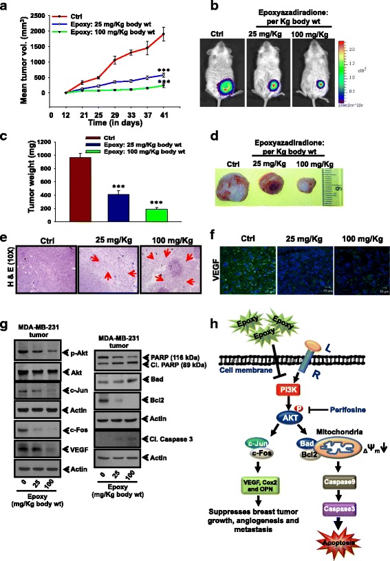
Fig. 7.

Epoxyazadiradione suppresses breast cancer growth and angiogenesis under in vivo conditions: a MDA-MB-231-Luc (2 × 10⁶) cells were injected orthotopically into NOD/SCID mice and then 25 and 100 mg/Kg body wt of epoxyazadiradione was injected intraperitoneally (i.p.) twice a week for 6 weeks. Tumor volumes were measured twice a week using Vernier Calipers, analyzed statistically and represented graphically (mean ± SEM, n = 5; ***, p < 0.0008 compared to untreated control tumor). b Photographs of bioluminescence imaging of representative tumor bearing NOD/SCID mice were analyzed using IVIS as indicated conditions. c and d Tumors were excised, photographed, weighed and analyzed statistically. Bar graph represents mean tumors weight (Mean ± SD, n = 5; ***, p < 0.0008). e Tumor sections derived from epoxyazadiradione treated or untreated mice were stained with H & E. Arrows indicate the necrotic area. f Tumor sections derived from epoxyazadiradione treated mice were analyzed for VEGF expression by confocal microscopy using anti-VEGF antibody. g Western blot analysis of p-Akt, c-Fos, c-Jun, VEGF and apoptosis associated molecules in the epoxyazadiradion treated tumor lysates. h Schematic representation of epoxyazadiradione regulated PI3K/Akt-mediated mitochondrial homeostasis, caspase-dependent apoptosis and attenuation of AP-1 activation, VEGF and MMP-9 expression and suppression of tumor growth and angiogenesis in breast cancer model
