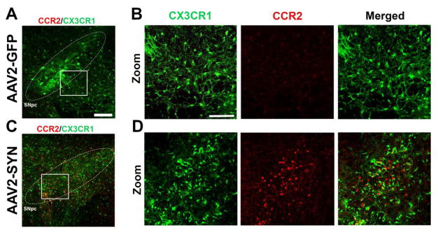
Figure 1. α-syn expression induces CCR2+ monocyte entry from the periphery.
(A) No detectable CCR2-RFP+ cells (CCR2, red) are observed in the SNpc (white circle, inset) of AAV2-GFP injected controls animals 4 weeks post-transduction in red/green mice. CX3CR1-GFP+ resident microglia (CX3CR1, green). Scale bar is 50 microns. (B) Zoom inset of AAV2-GFP injected controls. Single channels and merged images. Scale bar is 75 microns. (C) CCR2-RFP+ cells (CCR2, red) infiltrate from the periphery in the SNpc (white circle, inset) of AAV2-SYN injected animals 4 weeks post-transduction in red/green mice. (D) Zoom inset of AAV2-SYN injected animals. Single channel and merged images.

