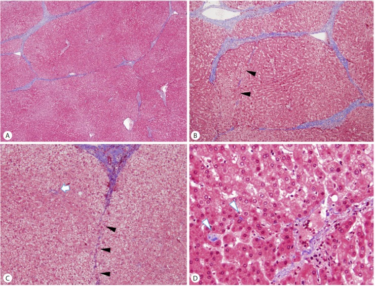
Figure 3.

An example of a cirrhosis with predominantly regressive pattern. (A) Cirrhosis with thin fibrous septa, corresponding to Laennec stage 4A. (B, C, D) Features of cirrhosis regression are seen in the field adjacent to (A), including perforated delicate septa (B, C; arrowheads), remnant portal tracts (C; arrow) and isolated collagen fibers (D; arrowheads). (Masson’s trichrome stain, original magnification ×40 (A, B), ×100 (C), ×200 (D))
