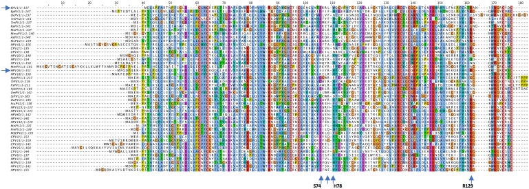
Fig 3. Multiple sequence alignment of the studied E6 protein set.
A protein multiple sequence alignment was performed using the MUSCLE program [103], and the output then entered into Jalview2 [109] to generate the ClustalX colorized image. The four upward-pointing arrows at the bottom of the alignment indicate the positions of HPV16 E6 amino acids that interact with position -3 of the E6AP LXXLL motif and BPV1 E6 interaction with PXN as shown in Fig 1. The two horizontal arrows on the left denote the positions of BPV1 E6 and HPV16 E6 in the alignment.

