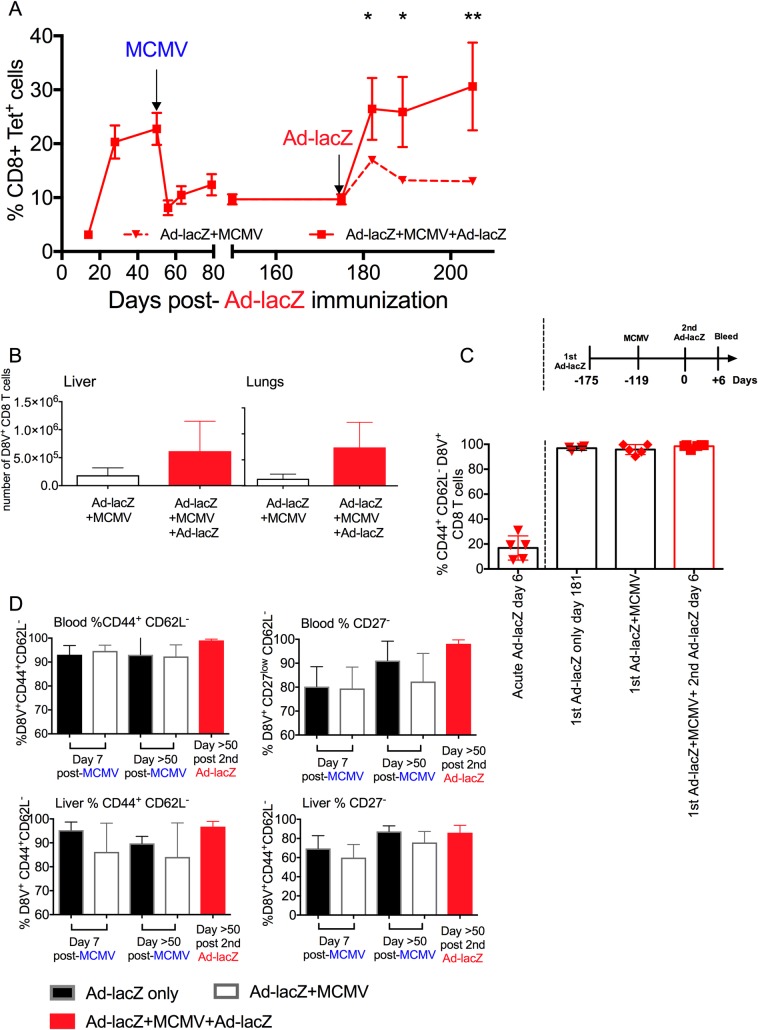
Fig 8. Effect of boosting with Ad-lacZ on the depleted responses.
(A) C57BL/6 mice were first immunized with Ad-lacZ and then >50 days later were infected with MCMV i.v. After >50 days post- MCMV infection, the mice were boosted with a second dose of Ad-lacZ i.v. The levels of CD8+D8V+ in the blood was measured by ex vivo tetramer staining after primary Ad-lacZ infection. The data shown are from one of two independent experiments (N = 3). (B) The distribution of the boosted cells in non-lymphoid organs were measured at day 50 after 2nd dose of Ad-LacZ. (C) The proportion of CD44+ CD62L- expression in ex vivo peripheral blood 6 days after primary Ad-lacZ immunization, and 6 days after second dose of Ad-lacZ. (N = 4–6 mice from 2 independent experiments). (D) The figures show the proportion of CD44+CD62L- (left column) and CD27-CD62L- (right column) expression in CD8+D8V+ cells from the blood (upper row) and liver (lower row) of Ad-lacZ only, Ad-lacZ+MCMV and Ad-lacZ+MCMV boosted with Ad-lacZ groups, as measured by ex-vivo staining. The data are from two or more independent experiments (N = 4–11). p values were measured by two-way Anova followed by Sidak’s multiple comparison test. *p<0.05, **p<0.005.

