Figure 1.
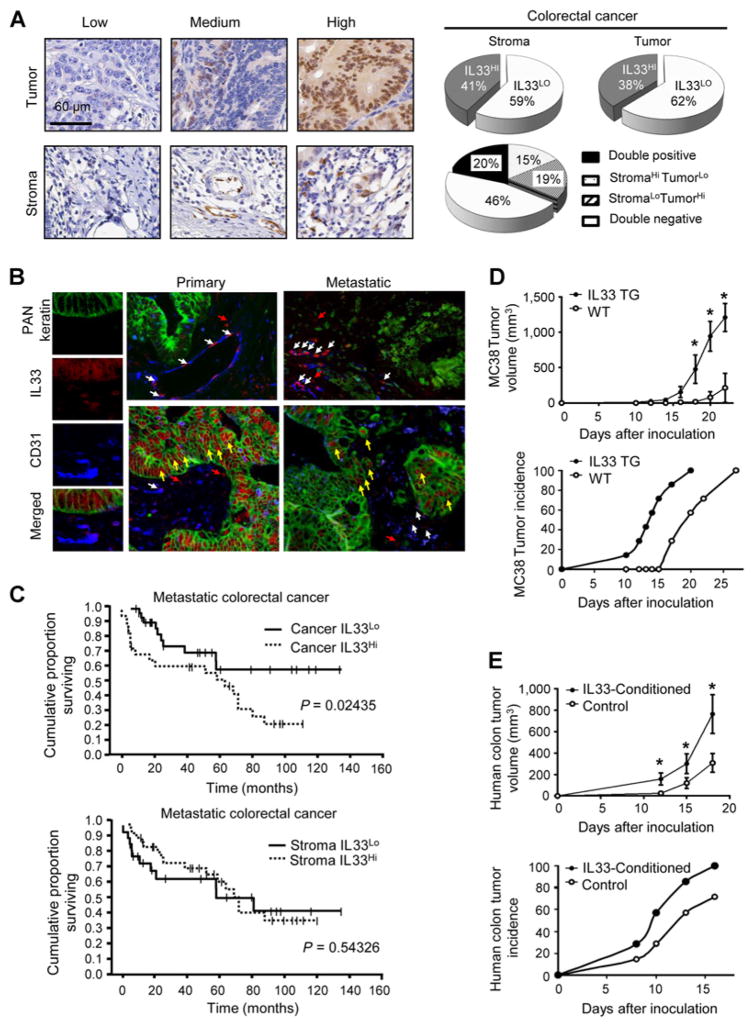
IL33 promotes colon tumorigenesis. A, IL33 expression was detected with conventional immunohistochemical staining in the human colon cancer tissues. The representative images of IL33 expression in colon cancer cells (Tumor) and stromal cells (Stroma) are shown. Scale bar, 60 μm. The proportion of IL33 expression in colon cancer cells and stromal cells in the colon cancer microenvironment is depicted (right, pie charts). B, IL33 expression was detected with multiplexed fluorescence staining in the human colon cancer tissues. The representative images show the expression of IL33 (red), CD31 (blue), PAN-Keratin (green). White arrows, nuclear IL33 localization in CD31+ vascular endothelial cells; yellow arrows, nuclear IL33 localization in Keratin+ tumor cells; red arrows, nuclear IL33 localization in CD31−Keratin− stromal cells. C, The association between the survival in patients with metastatic colon cancer and IL33 protein levels in tumor cells (top) and stromal cells (bottom). Survival functions were estimated by Kaplan–Meier methods and analyzed based on the H-score for tumor or stromal cell IL33 expression. D, MC38 cells (106) were subcutaneously injected into wild-type (WT) or IL33 transgenic (IL33 TG) mice. The tumor volume (top) and tumor incidence (bottom) were monitored. Results are expressed as the mean of tumor volume ± SEM; n = 7. *, P < 0.05. E, Human primary colorectal cancer cells (#1) were cultured with or without rhIL33 (0.1 μg/mL) for 24 hours. The cells (106) were subcutaneously injected into nude mice. The tumor volume (top) and tumor incidence (bottom) were monitored. Results are expressed as the mean of tumor volume ± SEM; n = 7, *, P < 0.05.

