Figure 5.
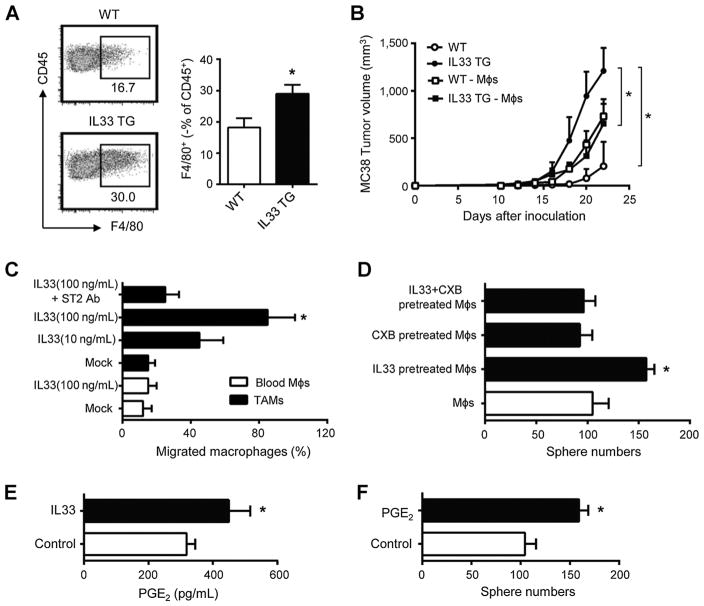
IL33 promotes colon cancer stemness via recruiting and stimulating macrophages. A, Tumor-associated macrophages in IL33 transgenic mice. MC38 cells (106) were subcutaneously injected into wild type and IL33 transgenic mice. Tumor-infiltrating immune cells were stained for CD45 and F4/80 and were analyzed by FACS. Results are shown as the mean of F4/80+ macrophages ± SEM in CD45+ cells in day 28 tumor tissues; n = 4; *, P < 0.05. B, Effects of macrophage depletion on MC38 tumor growth. MC38 cells (106) were subcutaneously injected into wild type or IL33 transgenic mice. Clodronate liposomes were intraperitoneally injected into the mice. Tumor growth was monitored. Results are expressed as the mean of tumor volume ± SEM. – Møs, macrophage depletion; n = 4 per group; *, P < 0.05. C, Macrophage migration toward IL33. CD14+CD45+ macrophages were enriched and sorted from colon cancer tissues or normal blood and subjected to the migration assay in the presence of IL33 and/or anti-ST2. Results are expressed as the mean percentage of migrated cells ± SEM. Møs, macrophages. TAM, tumor associated macrophages; n = 4; *, P < 0.05. D, Effects of IL33-treated macrophages on colon cancer sphere formation. Normal blood CD14+ macrophages were treated with IL33 in the presence or absence of celecoxib for 72 hours. Primary colon cancer cells were subject to sphere formation in the presence of these treated macrophages. Results are expressed as the mean of sphere numbers ± SEM; n = 4 per group; *, P < 0.05. E, Effects of IL33 on macrophage-derived PGE2.Normal blood CD14+ macrophages were treated with IL33 for 48 hours. PGE2 was detected in the culture supernatants by ELISA. Results are expressed as the mean values ± SEM; n = 4 per group; *, P < 0.05. F, Effects of PGE2 on colon cancer sphere formation. Primary colon cancer cells were subject to sphere assay in the presence of PGE2 (50 ng/mL). Results are expressed as the mean of sphere numbers ± SEM; n = 4 per group; *, P < 0.01.

