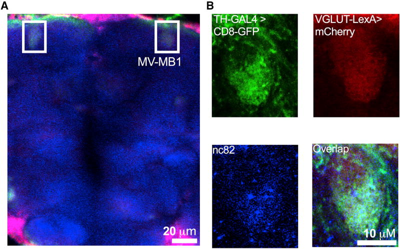
Figure 4. dVGLUT Localizes to Subpopulations of DA Nerve Terminals.
Fluorescent tags were co-expressed by dVGLUT-LexA (mCherry) and TH-GAL4 (mCD8::GFP) enhancer-driven expression drivers in WT back-ground.
(A) Representative image from a projected z series highlights co-localization of dVGLUT- and TH-promoter driven fluorescent tags in the DA terminal-rich MB-MV1 region (boxed in white; 20 µm scale bar; false color).
(B) Single-channel images zoomed in on MV-MB1 show TH-GAL4-driven mCD8::GFP (top left; green), VGLUT-LexA-driven mCherry (top right; red), and nc82 (labeling synaptic active zone marker Bruchpilot; bottom left; blue). Merged image (bottom right) shows co-localization of these fluorescent tags and nc82 labeling in DA terminal active zones. Fly strain: VGLUT-LexA/LexAOP-6x mCherry;TH-GAL4/UAS-mCD8::GFP. Data are representative of n > 3 experiments.
See also Figures S2 and S3.

