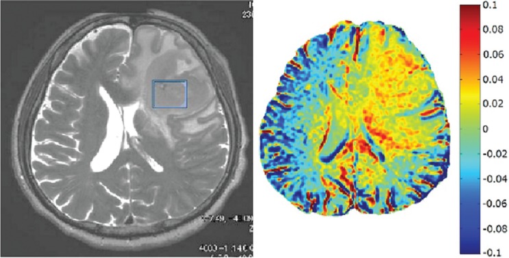
Fig. 7.
The source image and amide proton transfer (APT) image around 3.5 ppm obtained on a 71-year-old patient with malignant lymphoma. The malignant lymphoma lesion is shown in both source and APT images. Note that an elevated APT value of about 5% is observed in the tumor area as compared to the normal area, indicating greater malignancy of amide proton.

