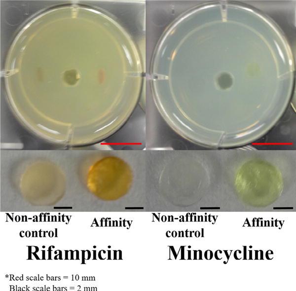
Fig. 2.

(Top images) In situ filling model with antibiotic filled disks after 52 h (RMP) and 45 h (MC) of filling. (Bottom images) Removed polymer disks after 52 h (RMP) and 45 h (MC) of filling. The dark orange (RMP) and yellow (MC) colors indicate the presence of the drug. The diameter of each well is 34.8 mm and all disks are 5 mm in diameter. Scale bars (bottom right corner of images) for wells are 10 mm (red) and scale bars for disks are 2 mm (black). (For interpretation of the references to colour in this figure legend, the reader is referred to the web version of this article.)
