
Figure 2. TGFBI−/− corneas show no aberrant changes.
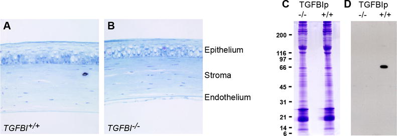
(A, B) No morphological changes are observed throughout the corneal in sections of (A) TGFBI+/+ and (B) TGFBI−/− corneas examined by light microscopy. Tissue sections are stained with toluidine blue; a 4x objective was used. (C) SDS-PAGE of TGFBI−/− corneas shows no major differences in protein expression when compared to control TGFBI+/+ corneas. (D) Western blotting using TGFBIp anti-serum confirms complete knockout in the TGFBI-deficient mouse eyes.