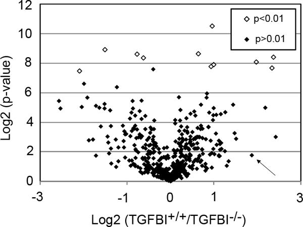
Figure 3. The ECM of TGFBI-deficient corneas is affected at the protein level.

The relative quantification of TGFBI+/+ and TGFBI−/− corneas using the iTRAQ methodology followed by LC-MS/MS resulted in 516 proteins being quantified in all three biological replicates of both the TGFBI+/+ and TGFBI−/− groups. The volcano plot shows the TGFBI+/+/TGFBI−/− ratio distribution of quantified proteins using the log2 scale compared to the log2 of the p-values. Using a p-value cut-off of 0.01, we identified 11 proteins (Table 1) that were differentially expressed in the TGFBI−/− cornea (empty dots). Six proteins (Type VI collagen, Type XII collagen, annexin, fibromodulin, prolargin and fibulin-5) known for their role in ECM arrangement were among the significantly mis-regulated proteins identified, suggesting that the ECM is altered in the TGFBI−/− cornea. The arrow indicates POSTN, a TGFBIp paralog, that shows a 1.9-fold upregulation on the log2 scale in the TGFBI−/− cornea; however, it was not significantly mis-regulated at p<0.01.
