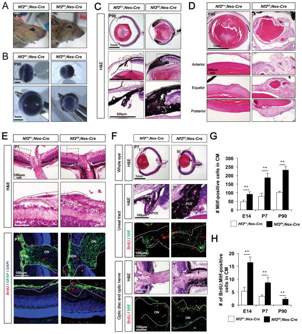
Figure 2. Loss of Nf2 in neuroepithelium results in the expansion of pigmented cells in the microphthalmic eyes.
Images of heads (A) and isolated eyes (B) of P90 Nf2f/+;Nes-Cre and Nf2f/f;Nes-Cre littermate mice. (C) Hamatoxylin and eosin (H&E) staining images of eye sections of P90 Nf2f/+;Nes-Cre and Nf2f/f;Nes-Cre littermate mice. The images in the bottom row are the magnified versions of the areas marked by dot-lines in the middle row. (D) H&E staining images of the lenses isolated from P90 Nf2f/+;Nes-Cre and Nf2f/f;Nes-Cre littermate mouse eyes (E) H&E staining images of the optic disc head in P7 littermate mouse eyes (top). Images in the second row shows magnified version of the areas surrounded by dot-line boxes in top row. Dot-lines outline the GCL and optic nerve. Astrocytes and proliferating cells in the optic disc head area are shown by immunostaining of GFAP and BrdU, respectively. ODH, optic disk head; ON, optic nerve. (F) Pigmented cells in the uveal tract (i and iii) and the optic nerve (ii and iv) of P7 Nf2f/+;Nes-Cre and Nf2f/f;Nes-Cre littermate mouse eyes sections in top row are magnified in the second and fourth rows with corresponding numbers. Immunostaining images in the third and fifth rows show proliferating cell incorporated with BrdU and pigmented cells expressing Mitf. (G) Quantification of Mitf-positive pigmented cells in the CM of Nf2f/+;Nes-Cre and Nf2f/f;Nes-Cre eyes at E14, P7 and P90. (H) Quantification of BrdU-positive cell population in Mitf-positive pigmented cells in Nf2f/+;Nes-Cre and Nf2f/f;Nes-Cre mouse eyes at E14, P7 and P90 (n = 6 from 3 independent litters). P-values (p) were obtained by Student’s two-tailed unpaired t-test (**, p < 0.01).

