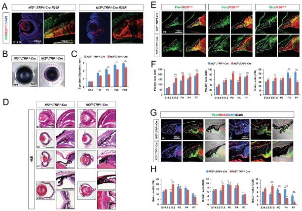
Figure 3. Nf2 loss in the CM and RPE resulted in the hyperplasia of the compartments.
(A) Nf2 immunoreactivity and R26lacZ (R26R) Cre recombinase activity reporter expression in the eyes of E14.5 Nf2f/+;TRP1-Cre;R26R and Nf2f/f;TRP1-Cre;R26R littermates. (B) Microscopic images of eyes isolated from P90 Nf2f/+;TRP1-Cre and Nf2f/f;TRP1-Cre littermates. (C) Quantification of eye sizes of Nf2f/+;TRP1-Cre and Nf2f/f;TRP1-Cre littermates at indicated ages (n = 6 from 3 independent litters). (D) H&E staining images of eye sections of Nf2f/+;TRP1-Cre and Nf2f/f;TRP1-Cre littermates at indicated ages. Images in right columns are magnified versions of dot-line box areas in left columns. (E) Sections of P0 Nf2f/+;TRP1-Cre;R26R and Nf2f/f;TRP1-Cre;R26R littermate mouse eyes were stained with antibodies against a pan-CM marker Pax6, an ICM and RPC marker Vsx2, and an OCM and RPE marker Otx2. CM areas are outlined by solid lines, and the borders between ICM and OCM are marked by dot-lines. (F) Quantification of Pax6-positive (left), Vsx2-positive (center), and Otx2-positive (right) CM cells in the staining images of Nf2f/+;TRP1-Cre and Nf2f/f;TRP1-Cre mouse eyes at indicated ages (n = 6 from 3 independent litters). (G) Nf2f/+;TRP1-Cre and Nf2f/f;TRP1-Cre eyes from P0 and P7 mice were analyzed to detect proliferating cells, which are positive to BrdU and Pax6 in the CM areas (outlined by solid lines). (H) Quantification of BrdU-positive cells in the whole CM (left), ICM (middle), and OCM (right) of Nf2f/+;TRP1-Cre and Nf2f/f;TRP1-Cre eyes (n = 6 from 3 independent litters). *, p < 0.05; **, p < 0.01; ***, p < 0.005.

