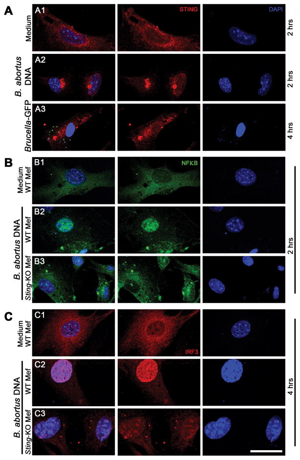
Figure 2. Brucella abortus DNA induces STING activation and translocation of NF-κB and IRF3.
MEFs from wild-type (WT) or STING KO were transfected with B. abortus DNA (1 μg/well, for 2 or 4 hrs as indicated) and cells from WT mice were infected with B. abortus S2308-GFP+ for 4 hrs (MOI 1000:1), fixed and subjected to immunofluorescence microscopy analysis of STING (A), NF-κB (B) or IRF3 (C). Pronounced translocation of STING was observed as aggregated speck formation in the perinuclear region 2 hrs after cells were transfected with bacterial DNA or 4 hrs after B. abortus S2308-GFP infection. STING-dependent-NFκB and -IRF3 activation was induced in wild-type MEFs by Brucella DNA transfection but not in KO cells. Antibody staining is shown in middle panels for STING (A), NF-κB (B) and IRF3 (C) and nuclei staining (DAPI) is shown in blue on the right panels. Left panels are merged images from those shown on middle and right panels. Data are representative of three independent experiments and three replicates in each experimental group. Size bar shown corresponds to 30 μm in all panels.

