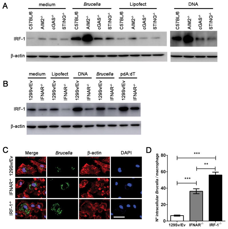
Figure 7. IRF-1 and type I IFN signaling are required to restrict Brucella replication in macrophages.
Macrophages derived from (A) C57BL/6, STING, cGAS and AIM2 KO or (B) 129Sv/Ev and IFNAR KO mice were infected with B. abortus (at MOI 100:1) or transfected with bacterial DNA (1μg/well) or poly dA:dT (1μg/well) encapsulated with lipofectamine or lipofectamine alone as control. Cell lysates were harvested 17 hrs after treatment and processing by western blot to determine the levels of IRF-1. (C) BMDMs derived from WT (129 Sv/Ev), IFNAR−/−, or IRF-1−/− mice were infected with Brucella-GFP (MOI 10:1) for 24 hrs and processed for fluorescent microscopy analysis. GFP-expressing bacteria are shown in green, phalloidin staining of the actin cytoskeleton for cell shape determination is shown in red, and DAPI (DNA) is shown in blue. Images show infected 129Sv/Ev, IFNAR−/−, or IRF-1−/− macrophages on top, middle and lower panels, respectively, as indicated on the left. Size bar shown on the lower-right panel in B corresponds to 30 μm in all panels. (D) The number of GFP-expressing bacteria was assessed for each cell, and 200 cells were analyzed for each mouse strain. Significant differences in average number of Brucella/cell comparing WT versus IFNAR and IRF-1 KO is denoted by *** (p<0.001) and IRF-1 versus IFNAR KO is denoted by ** (p<0.01 two-way ANOVA). Data are representative of three independent experiments and three replicates in each experimental group.

