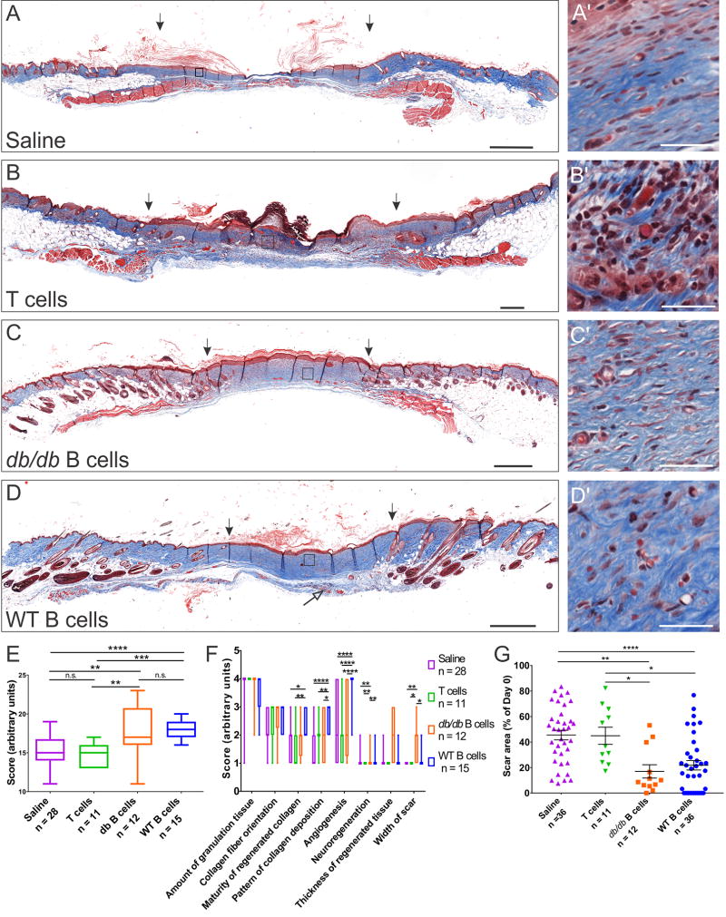
Figure 3. B cell treatment at the time of injury improves the regenerative outcome in an obese diabetic mouse model.
A–D Transverse sections through the wound bed at 34 days post-injury were stained with Masson’s Trichrome and evaluated for collagen deposition and maturity (blue). Scale bars, 500 µm. Areas enclosed in squares are shown at high magnification on the right (A’–D’). Scale bars, 40 µm. The thickness of the regenerated tissue is increased in wounds treated with B cells derived from either WT or diabetic animals, while the width of the scar at this time point was significantly reduced (arrows indicate the edges of the scar tissue). Nerve endings growing under and into the scar tissue were frequently observed in the wounds treated with B cells (D, open arrow). By contrast, T cell treatment led to a higher level of immune cell infiltration at the wound site and poor wound healing, with pronounced hyperkeratosis (B, B’). E. Blinded scoring of the scar sections based on aggregate histological criteria showed a significant advantage of wounds treated with B cells derived from either WT or db/db animals as compared to saline or T cell controls. F. Analysis of individual scoring criteria indicates that the B cell treatment is associated with improved deposition and maturation of collagen fibers, increased angiogenesis and neuroregeneration as well as a reduction in the width of the scar. G. Scar surface measurements at the end point of healing experiments show a significant reduction in the area of the scar in wounds treated with B cells at day 0. Data were pooled from 6 independent experiments. Significance was assessed using one-way (E, G) or two-way ANOVA (F), followed by Tukey’s multiple comparisons test. * p < 0.05; ** p < 0.01; *** p < 0.001; **** p < 0.0001.

